Apocrine
| Exocrine secretion |
| Merocrine or eccrine – by exocytosis |
| Apocrine – by membrane budding (loss of cytoplasm) |
| Holocrine – by membrane rupture |
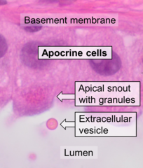
Apocrine (/ˈæpəkrɪn/)[1] is a term used to classify the mode of secretion of exocrine glands. In apocrine secretion, secretory cells accumulate material at their apical ends, often forming blebs or "snouts", and this material then buds off from the cells, forming extracellular vesicles. The secretory cells therefore lose part of their cytoplasm in the process of secretion.
An example of true apocrine glands is the mammary glands, responsible for secreting breast milk.[2] Apocrine glands are also found in the anogenital region and axillae.[3]
Apocrine secretion is less damaging to the gland than holocrine secretion (which destroys a cell) but more damaging than merocrine secretion (exocytosis).
-
Apocrine secretion
-
Apocrine gland
-
Histology of apocrine cells, H&E stain.
Apocrine metaplasia
[edit]
Apocrine metaplasia is a reversible transformation (metaplasia) of cells to an apocrine phenotype. It is common in the breast in the context of fibrocystic change. It is seen in women mostly over the age of 50 years. Metaplasia happens when there is an irritation to the breast (breast cyst). Apocrine-like cells form in a lining of developing microcysts, due to the pressure buildup within the lumen. The pressure build up is caused by secretions.[5] This type of metaplasia represents an exception to the common rule of metaplasia increasing the risk for developing cancer in that apocrine metaplasia doesn't increase the possibility of developing breast cancer.[6] Metaplastic apocrine cells belong to the category of oncocytes, which are a group characterized by abundant acidophilic, granular cytoplasm (from the Greek root onco-, which means mass, bulk).
Apocrine ductal carcinoma in situ
[edit]Apocrine ductal carcinoma in situ (ACDIS) is a very rare breast carcinoma which is regarded as a variant of the ductal carcinoma in situ breast tumors. ACDIS tumors have microscopic histopathology features that are similar to pure apocrine carcinoma of the breast tumors but differ from them in that they are completely localized, i.e. have not invaded nearby tissues or metastasized to distant tissues.[7]
Apocrine carcinoma
[edit]Apocrine carcinoma is a very rare form of female breast cancer. The rate of incidence varies from 0.5 to 4%.[8] Cytologically, the cells of apocrine carcinoma are relatively large, granular, and it has a prominent eosinophilic cytoplasm.[9] When apocrine carcinoma is tested as a “triple negative", it means that the cells of the patient cannot express the estrogen receptor, progesterone receptor, or HER2 receptor.[10]
References
[edit]- ^ "Apocrine | Meaning of Apocrine by Lexico". Lexico Dictionaries | English. Archived from the original on July 26, 2020.
- ^ Mescher AL, "Chapter 4. Epithelial Tissue" (Chapter). Mescher AL: Junqueira's Basic Histology: Text & Atlas, 12e: http://www.accessmedicine.com/content.aspx?aID=6180489.
- ^ Murphrey, Morgan B.; Safadi, Anthony O.; Vaidya, Tanvi (August 10, 2020). Histology, Apocrine Gland. StatPearls Publishing. PMID 29489220.
- ^ Image by Mikael Häggström, MD. Reference for findings: Carlos C. Diez Freire, M.D., Shahla Masood, M.D. "Apocrine metaplasia". Pathology Outlines.
{{cite web}}: CS1 maint: multiple names: authors list (link) Last author update: 28 May 2020. - ^ Dr Ayush Goel and Radswiki et al. Apocrine metaplasia of the breast.http://radiopaedia.org/articles/apocrine-metaplasia-of-the-breast
- ^ Wells, C A; El‐Ayat, G A (December 2007). "Non‐operative breast pathology: apocrine lesions". Journal of Clinical Pathology. 60 (12): 1313–1320. doi:10.1136/jcp.2006.040626. ISSN 0021-9746. PMC 2095572. PMID 18042688.
- ^ Quinn CM, D'Arcy C, Wells C (January 2022). "Apocrine lesions of the breast". Virchows Archiv. 480 (1): 177–189. doi:10.1007/s00428-021-03185-4. PMC 8983539. PMID 34537861.
- ^ Khandeparkar, Siddhi Gaurish Sinai, Sanjay D. Deshmukh, and Pallavi D. Bhayekar. "A rare case of apocrine carcinoma of the breast: Cytopathological and immunohistopathological study." Journal of Cytology/Indian Academy of Cytologists 31.2 (2014): 96.
- ^ "Final Diagnosis -- Case 209". Archived from the original on 2000-03-12. Retrieved 2014-11-03. (accessed November 3, 2014)
- ^ Potter, Michelle. "A[pcrome Breast Cancer - Johns Hopkins Kimmel Cancer Center". www.hopkinsmedicine.org.


